Surgical Options
Spine Surgery
Colorado Spine Partners emphasizes non-surgical treatment options for radicular pain for 8 weeks in advance of recommending spine surgery. But once non-surgical options fail to work, we believe in speeding the person back to activity with a minimally invasive spine surgery that can be done in day surgery so they are back home the same day.
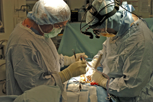 The surgeons at Colorado Spine Partners are fellowship-trained—the highest level of training in spine
surgery. They are also board-certified and concentrate their practice
exclusively on spine surgery. Their expertise includes minimally
invasive spine surgery, all common surgical problems in the neck
and low back, scoliosis care and surgery, as well as new innovations
including total disc replacement provided by Dr. Jantana. In addition, the surgeons at
Colorado Spine Partners are already referred some of the most complex spine
cases throughout the Denver region and have extensive experience
with cervical myelopathy, deformity surgery
and revision surgery. This experience translates into improved
patient care of all spinal problems.
The following is a list of common spine surgeries performed by Colorado Spine Partners
Common spine surgeries
Minimally Invasive Spine Surgery | Cervical Spine (Neck) | Lumbar Spine (Low Back) | Scoliosis | Spinal Fusion | Artificial disc
Minimally Invasive Spine Surgery
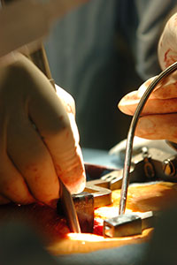
Colorado Spine Partners uses state of the art minimally invasive techniques and instrumentation to help patients recover in a shorter period of time and allow for a quicker return home. In minimally invasive spine surgery, a smaller incision is made, sometimes only a half-inch in length. The surgeon inserts special surgical instruments through these tiny incisions to access the damaged disc in the spine. For example, Colorado Spine Partners are proficient in the new Lateral Lumbar Interbody Fusion (XLIF) surgery to resolve leg or back pain from disc problems. Unlike traditional fusion surgery which requires a larger incision and hospital stay, XLIF is done through the patient’s side with a narrow probe and the patient can go home later the same day. Minimally invasive spine surgery requires extensive training and experience to master use of the tools, but there is tremendous benefit for the patient. The incision is shorter, resulting in less disruption to muscle and tissue as well as shorter hospital stay and quicker recovery time.
[Top]
Cervical Spine / Neck
Discectomy is the removal of the herniated portion of a disc to relieve the pressure on nearby nerves as they exit the spinal canal. Contrary to myths, the disc does not slip out of position like a watermelon seed. Instead, the disc is like a jelly donut, acting as the functional shock absorber between two bony vertebrae. An injury, damage from a lifting incident or a twist may cause the jelly center to break through the wall of the disc. When a disc herniates, the jelly center can press on nearby nerves. In the neck, this causes arm, shoulder, scapula and, in extreme cases, spinal cord compression.

Posterior Cervical Foraminotomy / Discectomy
For some herniated discs or bone spurs in the neck affecting only the nerve roots, a posterior discectomy and foraminotomy can be performed. This avoids spinal fusion, and with a microscope or a minimally invasive technique, can minimize recovery time and speed a patient back to work or activities.
Anterior Cervical Discectomy
Cervical is the medical term for "neck." Just as in a lumbar discectomy, the surgeon will remove a piece of damaged disc tissue in the neck area to relieve pressure on the spinal cord or nerve roots. In some cases, by removing a piece of the shock-absorbing disc that separates the two vertebrae, the structures may become less stable. Consequently, when the disc is removed, a surgeon may recommend "fusing" the vertebrae to prevent instability. A cervical discectomy is best left to surgeons who specialize in spine.
Corpectomy
A corpectomy is often performed for patients suffering from multiple levels of cervical stenosis with cord compression. The goal of a corpectomy is complete decompression of the spinal canal when stenosis encompasses more than just disc space and has moved into vertebral bodies.
Bone spurs forming toward the back of a vertebral body or the ligament behind vertebral bodies can cause the cervical spinal canal to narrow. Therefore, it may be necessary to remove one or more degenerating vertebrae and the discs above and below in order to decompress the spinal cord and nerve roots.
A corpectomy involves a vertical incision in the neck. The middle portion of the vertebra and its adjacent discs are removed to achieve decompression of the cervical spinal cord and nerve roots. A fusion accompanies a corpectomy surgery, using bone harvested from the patient's hip or from a bone bank. This bone graft is used to reconstruct the spine and provide stability.
Anterior Cervical Fusion
A fusion accompanies a anterior cervical discectomy or corpectomy. During fusion surgery, a disc Is removed, and the surgeon inserts a small wedge of bone between two vertebrae to restore disc space. Over time, the two vertebrae "fuse" together into a single solid structure. While this procedure limits movement and flexibility, it also helps relieve relieve neck pain.
Bone graft for the purpose of spinal fusion may be harvested from the patient's hip (autograft bone), from a cadaver bone (allograft bone), or from synthetic bone graft substitutes, which are currently being developed more extensively. Your surgeon will help you decide what is best for you.
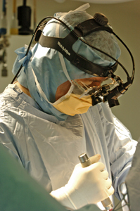
Laminoplasty
A laminoplasty, similar to a laminectomy, is
often performed on patients suffering from spinal stenosis in
the neck (narrowing of the cervical spinal canal). It is also
referred to as an "open-door
laminoplasty."
A laminoplasty creates more space for the spinal cord and roots.
The actual procedure involves cutting a “hinge” into
one side of the lamina and swinging it open like a door. It relieves
pressure on the spinal cord by increasing the diameter of the spinal
canal and room for the spinal cord. The surgery approach is through
the back of the neck. This procedure creates excellent spinal canal
decompression without the instability that may be created by multiple
level cervical laminectomies.
[Top]
Lumbar surgery/low back
Microdiscectomy / Minimally invasive discectomy
Discectomy is the removal of the herniated portion of a disc to relieve the pressure on nearby nerves as they exit the spinal canal. Contrary to myths, the disc does not slip out of position like a watermelon seed. Instead, the disc is like a jelly donut, acting as the functional shock absorber between two bony vertebrae. An injury, damage from a lifting incident, or a twist may cause the jelly center to break through the wall of the disc. When a disc herniates, the jelly center can press on nearby nerves. This causes back or leg pain when the hernation is in the low back, and arm pain if the disc is in the neck area (see "cervical spine/neck").
In a lumbar discectomy, the surgeon typically only removes the portion of the disc that is causing a problem, not the entire disc. If you have a herniated disc, keep in mind that a disc has a purpose. When you remove a disc, it may cause instability in the joint, and a surgeon may recommend a fusion to re-stabilize the area.
The surgeon can remove the damaged piece of disc through a traditional incision in the back. However, at Colorado Spine Partners, the surgeons typically use a microscope to minimize incision size, tissue trauma and recovery time. In addition, in some cases, minimally invasive discectomy can provide an even less invasive approach.
Depending on the nature of your disc problem, your surgeon will recommend the most appropriate type of surgery for you.
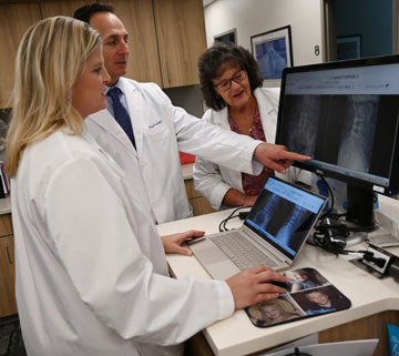 Anterior lumbar interbody fusion (ALIF)
In this type of spinal fusion surgery, back muscles and nerves remain undisturbed. The space between discs is fused by approaching the spine through the abdomen. This procedure is used when the spine is relatively stable, when there's a significant amount of disc space collapse, and in cases of one or two level degenerative disc disease. The surgeon will approach the abdomen through an incision (minilaparotomy) or by using an endoscope.
Posterior lumbar interbody fusion (PLIF)
This spinal fusion surgery is very similar to the anterior lumbar interbody fusion, except that the surgeon approaches the spine through the low back. This method is used when there is a greater amount of instability in the patient's spine. An advantage to this surgery is that it can also provide anterior fusion of the disc space without having a second incision.
Lumbar laminectomy
A laminectomy involves the removal of part or all of the bone covering the spinal canal. The purpose of this procedure can be to free nerve roots, remove a tumor, bone spur or to perform certain types of fusion procedures. Removing the lamina (laminectomy) is much like removing the cover on a fuse box to access the wiring. By removing the lamina, the surgeon gains access to the disc area and frees more space for the nerves inside.
A laminectomy in the lumbar spine is often used to treat recurrent disc herniations or where scar tissue is involved. Laminectomy may also be used in cases of spinal stenosis in which the entire canal is narrowed like a ring on a swollen finger, squeezing all of the nerve roots at that level of the spinal canal.
Lumbar disc replacement / artificial disc
In October 2004, the FDA approved total disc replacement (artificial disc), also called total disc arthroplasty. Some patients with degenerative disc disease who previously were candidates for spinal fusion may benefit from this motion-sparing technology.
The surgeons at Colorado Spine Partners are not only
trained in the implantation of the lumbar artificial disc, they are also
involved as a clinical trial site in the FDA's pending approval of a
cervical artificial disc. Surgeons at Colorado Spine Partners are committed to the
clinical expertise and technical precision that this new technology demands.
[Top]
Scoliosis and spinal deformity surgery 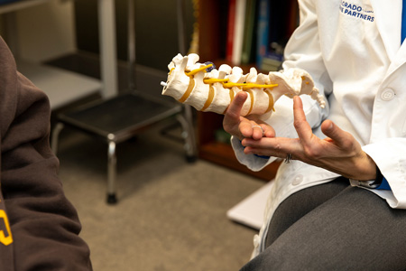
Through the placement of hooks, rods and screws, a spinal curve can be corrected and stabilized. A fusion often follows scoliosis surgery, in order to maintain the correction permanently.
Scoliosis is not the result of an injury and usually appears without cause. It can be inherited, and it usually affects more women than men. In the case of most spinal curves, the spine is not only bent but also twisted like a bent corkscrew. Some cases of scoliosis are not serious. Over time, if a curve worsens, surgery may be required to correct the curve and prevent pain and worsening deformity. In extreme cases, if the curve is not corrected, the spinal deformity can place pressure on internal organs, which can shorten a person's life expectancy.
During scoliosis surgery, the surgeon may
use special instruments that attach onto various vertebra segments.
These surgical rods are the adjusted to "de-rotate" the twisted and bent
corkscrew spine. Decades ago, Harrington Rods (the “first-generation”
of instrumentation) were used to surgically straighten the spine.
However, this technique did not untwist or correct the spine.
Today, there are “fourth-generation” techniques to
improve corrections, minimize levels fused and minimize the need
for post-operative bracing.
[Top]
Spinal fusion
Unusual movement at a vertebral segment will probably result in pain, especially if the person already has or displays symptoms of degenerative disc disease, fractures, scoliosis or a weak spine. This movement may require a discectomy and subsequently a lumbar interbody fusion. Anterior and posterior fusion techniques can be performed in the neck and the low back.
Not all
patients who have spinal problems need spine surgery. They can be managed
with microscopic decompression or minimally invasive techniques. Spinal
fusion is reserved for patients who have spinal instability, spinal deformity
or painful degenerative pain. Obviously, this is only after a patient
has failed all conservative measures. In fusion surgeries, the goal is
to cause bone graft to grow between two vertebrae and stop the motion
at a particular segment by adding bone graft to it. This results in one
long bone rather than two separate vertebrae. Anterior and posterior
lumbar fusions may be done separately or can be used together for the
most severe problems of the cervical (neck), thoracic (chest level) and
lumbar spine (low back). Your spinal surgeon will help you decide which
technique is right for you.
[Top]
Artificial disc implantation
One of the most anticipated advances in spine surgery over the past 20 years was the arrival of the artificial disc. The first artificial disc in the United States received formal approval by the Food and Drug Administration (FDA) for widespread use in the United States on October 26, 2004. While this technology is still somewhat new to the U.S., artificial discs have been in use in Europe for more than 20 years.
It is important to remember that this technology is still evolving with new implants continually in development. Your spine surgeon is the best resource to discuss if it is appropriate for you, and what model of artificial disc is best suited for your case.
For example, there are several models of artificial discs approved by the FDA for use in the United States and that number is expected to grow as new models emerge on the scene and surgeons become trained in their use. Each disc is designed for use either in the low back (lumbar area) or neck (cervical area).
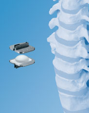
Prodisc-C © by Synthes
In addition, other artificial disc models are available on a limited basis through “clinical trials” where a patient agrees to be part of clinical study designed to measure the success of a new disc model, which will be part of an FDA study. Patients who participate in a clinical trial can gain access to the most current technology, even though it has yet to gain FDA approval for use in the U.S.
At Colorado Spine Partners, Dr. Sanjay Jatana uses the ProDisc-C for the cervical spine and the ProDisc-L for the lumbar spine.
An Alternative to Fusion Surgery
The artificial disc concept is intended to be an alternative for spinal fusion surgery. Each year in the U.S., more than 200,000 spinal fusion surgeries are performed to relieve excruciating pain caused by damaged discs in the low back and neck areas.
During a fusion procedure, the damaged disc is typically replaced with bone from a patient’s hip or from a bone bank. Fusion surgery causes two vertebrae to become locked in place, putting additional stress on discs above and below the fusion site, which restricts movement and can lead to further disc herniation with the discs above and below the degenerated disc. An artificial disc replacement is intended to duplicate the function level of a normal, healthy disc and retain motion in the spine.
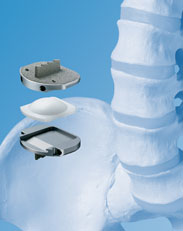
Prodisc-L © by Synthes
When a natural disc herniates or becomes badly degenerated, it loses its shock-absorbing ability, which can narrow the space between vertebrae. In fusion surgery, the damaged disc isn’t repaired but rather is removed and replaced with bone that restores the space between the vertebrae. However, this bone locks the vertebrae into place, which can then damage other discs above and below.
A common aspect of all artificial discs is that they are designed to retain the natural movement in the spine by duplicating the rotational function of the discs Mother Nature gave us at birth. Most artificial disc designs have plates that attach to the vertebrae and a rotational component that fits between these fixation plates. These components are typically designed to withstand stress and rotational forces over long periods of time. Still, like any manmade material, they can be affected by wear and tear.
Benefits
Some of the main benefits of the artificial disc parallel that of knee replacement and hip replacement. This can include the following benefits:
- An artificial disc in the neck or back, in principle, is designed to retain motion in that particular segment of the spine.
- It prevents degeneration of disc levels above and below the affected disc
- There is no bone graft required
- There can be a quicker recovery and return to work or activity
- It can be a less invasive and less painful surgery than a fusion
- There can be less blood loss during surgery
Lumbar vs. Cervical Artificial Discs
Because of the weight of the body and the rotational stress that the trunk places on discs in the low back (lumbar) area, more stress is placed on artificial discs in the lumbar area than in the neck (cervical) area, which only supports the weight of the head.
A second issue relates to the ease of the artificial disc surgery and any necessary revision surgery to replace a worn out artificial disc. Because the surgeon must access the front of the spine, an incision is made in the abdomen for lumbar discs and in the front of the neck for cervical discs. Generally speaking, many spine surgeons believe access to the cervical discs can be easier than the lumbar discs.
 Other issues to consider
When treating knee and hip replacement patients, orthopedic surgeons try to postpone the implantation of an artificial joint until a patient is at least 50 years old so that they do not outlive their artificial joint, which typically lasts anywhere from 15 to 20 years. Revision surgery, which may be necessary to replace a worn-out artificial joint, can be complex.
This is also a concern with the artificial disc. Unlike knee and hip replacement patients who are typically in their 50s or 60s, many patients can benefit from artificial disc technology at a much younger age — in their 20s or 30s. Therefore, the implantation of an artificial disc in younger patients can raise a surgeon’s concern about the potential life span of the artificial disc in the spine and the need for revision surgery to replace a worn-out artificial disc, which can be complex.
In summary, some spine surgeons may be cautious about the use of artificial discs for the following reasons:
- Wear and tear on artificial joints cant require revision surgery in 10 to 20 years that can be extremely complex.
- Most artificial disc implants only address rotational forces, not the up and down shock absorbing function of the natural disc.
- Overweight people can wear out a lumbar disc prematurely.
- New artificial discs are continually in development, however FDA approval is a lengthy process.
- There are not many 20-year-long studies that show the long-term effects of wear and tear on artificial disc implants.
Generally, the technology is very promising. Your spine surgeon can provide information if your problem can be addressed with this technology.
[Top]
