Diagnostic Testing
Evaluation of back and neck pain requires a physician who is experienced in diagnosing spinal conditions. The work-up begins with a detailed history and physical examination. Your medical history helps the doctor understand your back and neck pain and the influence of your lifestyle on your spine.
During the physical exam, your physician will try to pinpoint the source of pain. Simple tests for flexibility and muscle strength may also be conducted. Diagnostic tests may be ordered to confirm the location and source of your pain.
 Diagnostics may include:
-
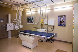
X-rays are usually the first step in diagnostic testing methods. X-rays show bones and the spaces between the bones.
-
MRI (magnetic resonance imaging) uses a magnetic field and radio waves to generate highly detailed pictures of the inside of your body. Because X-rays only show bones, MRIs are needed to see soft tissues like spinal discs. These images help your doctor provide a more accurate diagnosis. MRIs are very safe and usually pain-free.
-
CT scan/myelogram - A CT scan is similar to an MRI because it provides additional diagnostic information about the internal structures of the spine. A myelogram is used to diagnose a bulging disc, tumor or changes in the bones surrounding the spinal cord or nerves. A local anesthetic is injected into your low back to numb the area. A lumbar puncture (spinal tap) is then performed. A dye is injected into the spinal canal to reveal where problems lie.
-
Electrodiagnostics - Electrical testing of the nerves and spinal cord may be performed as part of our diagnostic workups. These tests, called electromyography (EMG) or somato sensory evoked potentials (SSEP), assist your physician in understanding how your nerves or spinal cord are affected by your condition.
Bone Scan - Bone imaging is used to detect infection, malignancy, fractures and arthritis in any part of the skeleton. Bone scans are also used for detecting lesions for biopsy or excision.
-
Discography - Discography is used to determine the internal structure of your disc. It is performed with a local anesthetic by injecting dye into the disc under X-ray guidance. An X-ray or CT scan is performed to determine if the disc’s structure is normal or abnormal and if the injection causes pain. A benefit of a discogram is that it enables the spine surgeon to determine the disc level that is causing pain. This ensures that surgery will be more successful by reducing the risk of operating on the wrong disc.
-
Injections - Pain-relieving injections can act as a bridge to physical therapy by relieving back pain and providing the physician with important information about your problem.